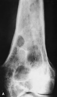
A 40 year old male, cattle farmer, presenting with pain in leg over 1 year. No history of significant trauma, infection or any other swelling in the body. Can u identify the pathology?
Discussion:
Musculoskeletal hydatid infections are a very rare form of Echinococcosis/hydatidosis.
Clinical presentation
Patients usually present with slow growing swelling with or without pain.
Location
They can present almost anywhere, but most common locations are:
- vertebrae and para-vertebral soft tissue
- pelvis
- femur and soft tissue of lower limb
- tibia
Radiographic features
Ultrasound
Ultrasound may depict lesions in the soft tissue which can be solitary/multiple unilocular/multilocular/complex cystic lesions and/or atypical solid hypoechoic lesion.
Radiograph
- may show expansile lytic lesion in the involved bone which can be unilocular or multilocular with coarse trabeculae
- thinning of cortex
- adjacent soft tissue swelling may be seen due to direct extension from the bone or co-existing multiple soft tissue lesions
CT
Better delineates expansile unilocular or multilocular lytic lesion and demonstrates any soft tissue extension. Co-existing soft tissue multilocular cysts may be seen.
MRI
- T1: hypointense cyst
- may show low-intensity rim
- T1 C+: shows wall enhancement
- T2: hyperintense cyst (uni/multilocular) with a low-intensity rim
- water lilly sign may be seen


Comments
Post a Comment